1
2
3
4
5
6
7
8
9
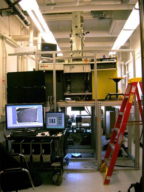
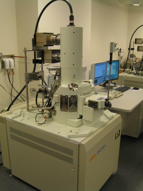
Reid Lab: T.E.M.C.A., may be the worlds fastest electron microscope, capable of continuously generating images at rates as high as 15 Mpixels/s. Many times faster than commercially available systems, it was fabricated for a fraction of the price. The Neurotechnology facility played a role in nearly every stage of this project.
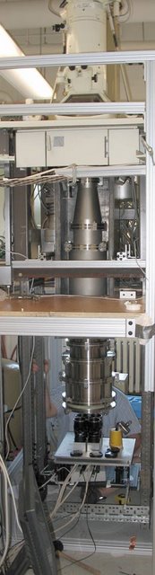
Reid Lab: Unlike a traditional T.E.M. in which an image is formed at the level of the film chamber (white rectangular flanked by squares), a large vacuum chamber allows the electron image to expand, much like the magnified image created by a movie projector. Steel and lead shielding is removed for photo.
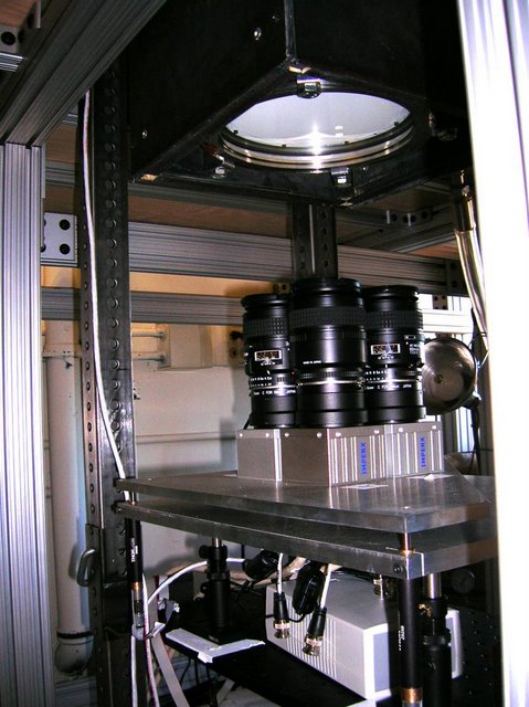
Reid Lab: A large phosphor screen at the end of the path is imaged by four overlapping 11 Mpixel CCD cameras.
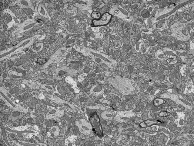
Reid Lab: An early image taken from the microscope.
Lichtman Lab: Scanning electron microscopes will play a large role in fulfilling the goals of the Connectome Project. We have provided software and hardware support; occasionally assisting the manufacturers with custom installations.
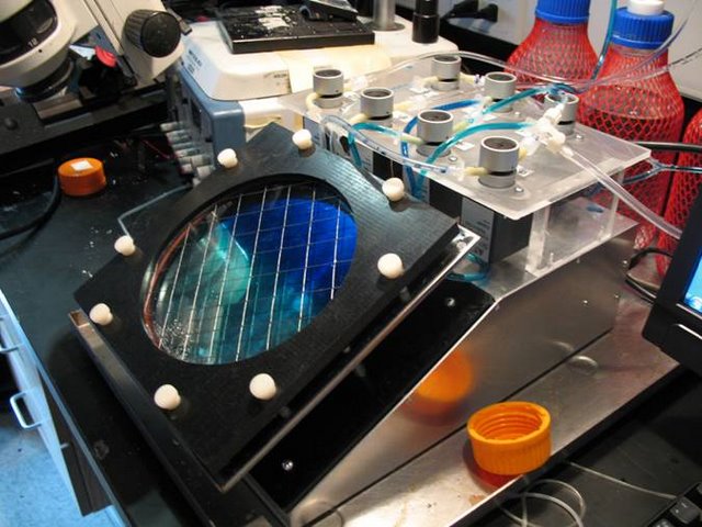
Lichtman Lab: In order to reproducibly stain large volumes of tissue sectioned by the A.T.L.U.M., we designed and fabricated this fully automated device. It provides temperature controlled staining with up to two solutions and a rinse. During prototyping, solution exchange was monitored with dyes.
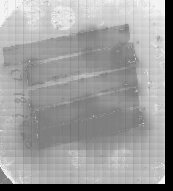
Lichtman Lab: The goals of the Connectome Project often demand features that were not implemented by microscope manufacturers. Shown here is an image of a four inch silicon wafer holding five strips of A.T.L.U.M. tape. In order to attain such a low power image, we created a custom microscope interface and image acquisition system.
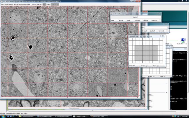
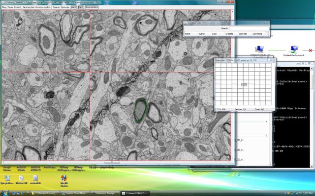
Lichtman Lab: Working with Duncan Mak, we created software capable of visualizing nearly boundless amounts of data. While many excellent commercial programs exist (Photoshop, Image J, etc.) most can not handle the 100’s of Gigabytes of data that are required by the Connectome. Red lines delineate individual overlapping 20Mpixel images that are part of a much larger sheet.
Lichtman Lab: The same data shown at higher magnification. The software also allows for searchable markup (green contour). We have tested its performance with image sheets as large as 1.5 Terabytes